Medical Cross Section Coloring: Techniques for Anatomical Detail
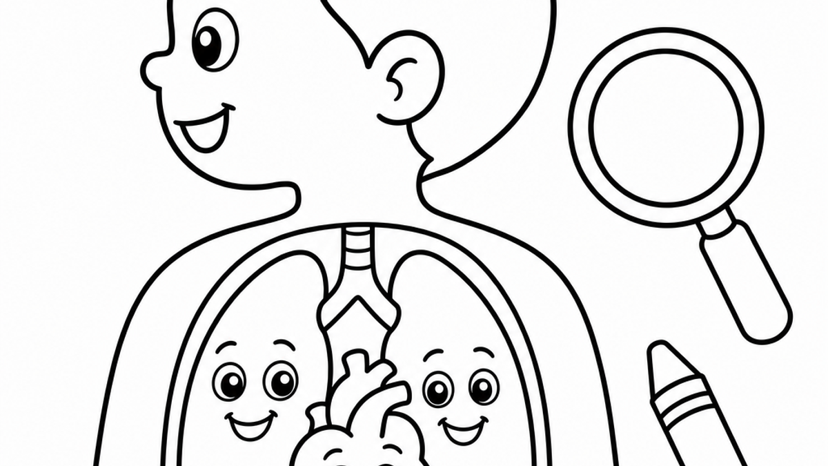
Anatomical Cross-Section Coloring for Medical Detail
A nursing student at a kitchen table at 11 p.m., quizzing herself on the layers of the epidermis with a pack of colored pencils instead of flashcards. A homeschool parent trying to explain why the heart has four chambers without just pointing at a photo and hoping for the best. A kid who wants to know what's actually inside a knee. Cross-section coloring pages turn out to be the right tool for all three, just at very different levels of detail.
This isn't the same category as a unicorn or a fire truck page. A cross-section diagram asks you to notice structure, not just fill in shapes. That's exactly why it works for studying.
What Is a Cross-Section Diagram in Anatomy?
A cross-section diagram shows what a structure looks like if you sliced straight through it. Instead of the outside of a heart, you see the chambers, valves, and vessel walls stacked and nested inside. Instead of a leg, you see skin, fat, muscle, bone, all the layers a scalpel or an MRI slice would reveal.
In medical illustration, this view matters because the body doesn't organize itself the way a textbook paragraph does. Structures sit on top of each other, wrap around each other, share walls. A cross-section is the only way to show that relationship clearly on a flat page.
Coloring one forces you to track where one layer stops and the next starts. That's the whole exercise, really. You can't shade the dermis the wrong color without noticing it butts right up against the epidermis above and the subcutaneous fat below.
Anatomy Coloring Pages for Medical Students and Nursing Students
Anatomy coloring books have been a study tool in medical and nursing programs for decades, which says something, since med students generally have access to every studying technology money can buy and still reach for pencils.
The logic holds up. Coloring a structure means looking at it closely enough to decide where its boundary is, which is a different kind of attention than reading a caption and nodding. You're not just recognizing the kidney. You're deciding where the cortex ends and the medulla begins, then committing to that line in pencil.
A few ways nursing and med students actually use these pages:
- Coloring the same structure from a few different diagrams to compare labeling conventions
- Using one consistent color per tissue type across a whole study session (all muscle red, all connective tissue yellow) to build pattern recognition
- Color-coding a diagram first, then covering the labels and re-quizzing from memory
We'll say the honest thing here: we can't promise coloring improves exam scores, and we haven't seen a controlled study that measures it directly. What we can say is that active, hands-on review tasks are a well-established complement to reading, and tracing structure by color is one low-cost way to do that.
Body Layer Coloring Pages: Skin, Muscle, and Organ Cross-Sections
Layers are where cross-section coloring earns its keep. A skin layer cross-section page typically shows the epidermis, dermis, and hypodermis stacked with hair follicles and sweat glands threaded through. A muscle layer anatomy coloring page usually shows the progression from skin down through fascia to the muscle fibers themselves, sometimes with a cross-section of the muscle bundle itself, fibers grouped into fascicles.
Here's where value and intensity actually matter, not just as art class vocabulary but as a study trick. The Getty Museum's framework on the elements of art describes value as how light or dark a color is and intensity as how pure or diluted it is. Using a consistent value scale, say, lighter shades for superficial layers and darker shades as you go deeper, turns the page into a visual key you can read at a glance instead of a flat wash of random colors.
Organ cross-section worksheets (kidney, heart, lung) work the same way. Color the outer capsule light, the inner structures darker, and the page starts explaining itself before you even get to the labels.
Medical Cross-Section Coloring Pages for Kids
For younger kids, we're not talking about fascicles and glomeruli. We're talking about a simplified skeleton, a friendly heart with big visible chambers, a stomach and intestines drawn like a road map rather than a photograph.
Skeletal anatomy coloring pages for kids work well because bones are already shape-based. Kids can color the skull, ribs, and leg bones as distinct puzzle pieces without needing to understand marrow or density. It's basically a body-shaped jigsaw with crayons.
Educational anatomy coloring pages for teens can step up the detail without going full medical-textbook. A teen in middle school biology can handle a labeled heart with the four chambers and major vessels, or a simplified brain diagram showing the cerebrum, cerebellum, and brain stem, without needing nursing-school precision.
A quick gut check on accuracy: a cross-section coloring page is a teaching illustration, not a diagnostic image. It's simplified on purpose, the way a subway map simplifies a city. That's a feature for a 7-year-old and a reasonable starting point for a teen, but a nursing student should always check a detailed page against a current anatomy atlas before treating it as exam-ready.
Small hands benefit from this kind of focused, contained coloring task too. The National Association for the Education of Young Children notes that activities like drawing, coloring, and tracing build the fine motor control kids use for things like handwriting later on. A detailed cross-section page, with its smaller regions and tighter lines, gives slightly older kids more of that fine motor challenge than a big bold animal page does. If a child in your house or classroom does better with occupational therapy style fine motor practice, our occupational therapy coloring sheets are built with that same small-muscle focus in mind.
Physiology Coloring Pages for Homeschool Biology
Homeschool biology is where cross-section coloring gets to do its best work, mostly because there's room in the schedule for a worksheet that takes twenty unhurried minutes instead of five rushed ones.
A few ways it fits into a homeschool biology unit:
- Pair a digestive system cross-section with a unit on nutrition, coloring each organ as you cover it that week
- Use a muscle layer page alongside a unit on movement and exercise
- Color a skin cross-section the same week you talk about sun safety, temperature regulation, or the five senses
It also works as an assessment that doesn't feel like a test. A kid who can correctly color and label a heart diagram from memory has demonstrated something real, without anyone handing out a scantron sheet.
This fits naturally into the kind of screen-free, hands-on stretch of the day that a lot of homeschool routines are built around in the first place. If your family already sets aside device-free blocks, a detailed worksheet like this slots right in without anyone needing to negotiate.
Frequently Asked Questions
What age is appropriate for anatomy coloring pages?
Simplified versions (big bony skeleton, friendly heart, basic digestive tract) work from around age 5 or 6. More detailed cross-sections with labels and smaller regions suit kids 9 and up, and the most detailed versions are really built for teens and adults.
Why do nursing students use coloring books to study anatomy?
Coloring forces close attention to boundaries between structures, which is useful when a lot of anatomy study is otherwise passive reading. It's a hands-on complement to a textbook, not a replacement for one.
How accurate are anatomical coloring pages for medical study?
They're simplified teaching illustrations, similar to a textbook diagram rather than a clinical scan. They're a solid study aid, but nursing and medical students should cross-check detailed pages against a current anatomy atlas before an exam.
Are cross section coloring pages good for homeschool biology?
Yes, as a hands-on supplement alongside reading and discussion. They work well for review and for low-pressure self-assessment, not as a stand-alone curriculum.
How to use coloring pages to teach kids about the body?
Start simple (skeleton, heart, basic organs), name each part out loud as you color it, and let the child ask questions about what each piece does. Save the more detailed, labeled versions for when they're a bit older.
Do anatomy coloring books help with memorization?
Many students and teachers find that active tasks like coloring and labeling support memorization better than reading alone, though individual results vary and we haven't seen a controlled study measuring coloring specifically against other review methods.
What is a cross section diagram in anatomy?
It's an illustration showing what a structure looks like if sliced straight through, revealing internal layers and chambers that an outside view can't show.
If your classroom or kitchen table needs a cross-section page on a specific structure this week, or a quieter stretch of quiet classroom coloring activities for review time, our generator can put together a printable page on exactly what you're studying in about two minutes.
The Chunky Crayon Editorial Team
Chunky Crayon editorial
The Chunky Crayon team publishes playful, practical and source-aware ideas for creative family time. Developmental or wellbeing claims are general education, not clinical or educational assessment.